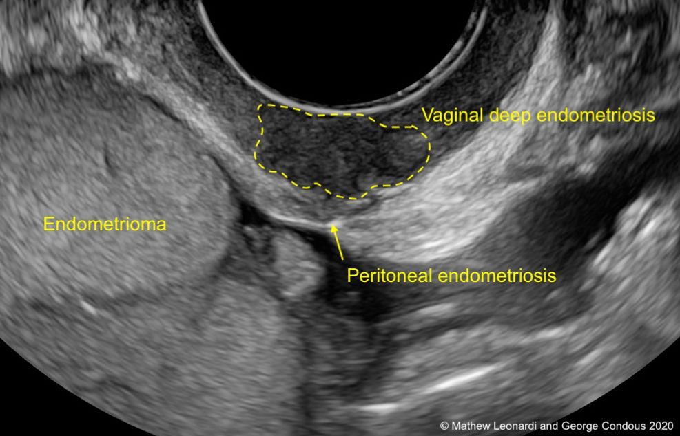
Endometriosis Ultrasound Ovary | The pathogenesis of intraovarian endometriomas may be different than that of endometriosis that occurs as superficial peritoneal implants. Looking for endometriosis involves looking not just at the uterus and ovaries but also at the bladder, the ligaments behind the uterus, the vaginal wall and the bowel. Transverse transvaginal scan of the uterus. Endometriomas are usually readily detectable by ultrasound. However, recent literature shows that in expert hands it can present a similar sensitivity to mri in the detection of intestinal endometriosis 35.
Endometriosis is defined as the presence of endometrial glands and stroma outside the uterine cavity 2 . There is minimal free fluid in the pelvis. An ultrasound imaging scan is a simple and fast way for your doctor to see inside your pelvis to assess your uterus, ovaries, and fallopian tubes if they. 4) (appearing as multiple cysts separated by septations) and is often multiple or bilateral. A transvaginal ultrasound scan is a safe and straightforward procedure that uses ultrasound waves to produce images of the womb (uterus), fallopian tubes, ovaries, and in centres specialising in endometriosis, other structures including the bladder and bowels.
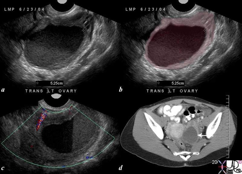
Women may also have endometrial cells growing in more distant areas of the body. Transvaginal ultrasound has been shown to be an accurate and reliable diagnostic tool in detecting ovarian endometrioma and deep infiltrating endometriosis. Ovarian remnants occur when the ovary is bluntly dissected from the pelvic sidewall when it is adhered or scarred down to the pelvic sidewall. I have been having symptoms of endo for a while now, and finally got the courage to go to the doctor about it. A transvaginal ultrasound scan is a safe and straightforward procedure that uses ultrasound waves to produce images of the womb (uterus), fallopian tubes, ovaries, and in centres specialising in endometriosis, other structures including the bladder and bowels. They are readily diagnosed on ultrasound, with most demonstrating classical radiographic features. This old blood has the appearance of thin chocolate or motor oil. The pathogenesis of intraovarian endometriomas may be different than that of endometriosis that occurs as superficial peritoneal implants. The most significant ovarian involvement is an endometrioma. Most commonly due to severe endometriosis/deep infiltration endometriosis, but can also be secondary to pelvic inflammatory disease. If the endometrial tissue is within an ovary, that ovary will fill with blood. 14 in the presence of endometriosis, the fallopian tubes may be impaired resulting in a hydrosalpinx or hematosalpinx. 3 found that only 51 % of the endometriomas were unilocular cysts with ground glass echogenicity of the cyst fluid.
Endometriosis is a painful condition in which tissue that normally lines your uterus (endometrial tissue) grows in other parts of your pelvis, such as your ovaries or fallopian tubes. The pathogenesis of intraovarian endometriomas may be different than that of endometriosis that occurs as superficial peritoneal implants. My gp was very sympathetic, she did a pelvic exam, swabs, and a urine test which all came back fine. 4) (appearing as multiple cysts separated by septations) and is often multiple or bilateral. The visualized myometrium is homogenous in appearance.
Ovarian endometriosis may be unilocular or multilocular (fig. An ultrasound imaging scan is a simple and fast way for your doctor to see inside your pelvis to assess your uterus, ovaries, and fallopian tubes if they. Ovarian remnants occur when the ovary is bluntly dissected from the pelvic sidewall when it is adhered or scarred down to the pelvic sidewall. This scarring is usually the result of endometriosis which causes inflammation, and the result of that inflammation is the formation of adhesions. Ultrasound imaging for ovarian and deep infiltrating endometriosis the main challenges of imaging for endometriosis are the detection of nonovarian disease and the evaluation of the extension of the disease into pelvic structures. They are also called chocolate cysts of the ovary. Women may also have endometrial cells growing in more distant areas of the body. During the ultrasound scan, you will be asked to lie on your back. I have been having symptoms of endo for a while now, and finally got the courage to go to the doctor about it. An accurate ultrasound diagnosis, which describes and documents the location and extent of endometriosis, improves patient care by allowing better preoperative surgical planning. Most commonly due to severe endometriosis/deep infiltration endometriosis, but can also be secondary to pelvic inflammatory disease. These adhesions can often be very dense and difficult to. It is mainly found in the abdominal cavity, most commonly on the surface of the ovaries.
Ultrasound imaging for ovarian and deep infiltrating endometriosis the main challenges of imaging for endometriosis are the detection of nonovarian disease and the evaluation of the extension of the disease into pelvic structures. This old blood has the appearance of thin chocolate or motor oil. Ovarian endometriosis may be unilocular or multilocular (fig. Transvaginal ultrasound has been shown to be an accurate and reliable diagnostic tool in detecting ovarian endometrioma and deep infiltrating endometriosis. Most endometriomas are located within the ovary.

Endometriosis most commonly involves your ovaries, fallopian tubes and the tissue lining your pelvis. These adhesions can often be very dense and difficult to. The implantation of endometrial tissue in the peritoneal cavity through retrograde menstruation is the most accepted theory of endometriosis but its etiology is still poorly understood 3 . Endometriomas occur when surface ovarian endometriosis lesions start to push into or invaginate into the ovary and then close over the top, forming a cystic mass lined by endometriosis tissue. Transverse transvaginal scan of the uterus. My endo lead to pelvic floor dysfunction and vulvodynia, so the combination of. I have been having symptoms of endo for a while now, and finally got the courage to go to the doctor about it. An ultrasound imaging scan is a simple and fast way for your doctor to see inside your pelvis to assess your uterus, ovaries, and fallopian tubes if they. A transvaginal ultrasound scan is a safe and straightforward procedure that uses ultrasound waves to produce images of the womb (uterus), fallopian tubes, ovaries, and in centres specialising in endometriosis, other structures including the bladder and bowels. Endometriosis is defined as the presence of endometrial tissue outside the uterine cavity. My gp was very sympathetic, she did a pelvic exam, swabs, and a urine test which all came back fine. They are also called chocolate cysts of the ovary. Unless endometriosis forms cysts on the ovaries (endometriomas) it is therefore not picked up with a traditional gynaecological ultrasound.
These adhesions can often be very dense and difficult to endometriosis ultrasound. This scarring is usually the result of endometriosis which causes inflammation, and the result of that inflammation is the formation of adhesions.
Endometriosis Ultrasound Ovary: It is mainly found in the abdominal cavity, most commonly on the surface of the ovaries.


0 Please Share a Your Opinion.:
Post a Comment